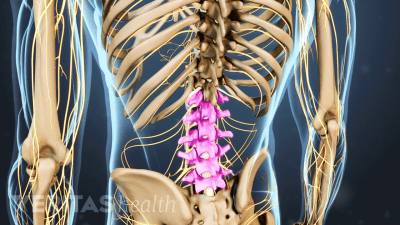
There are 5 pairs of spinal nerves in the lumbar spine, labeled L1 to L5.
Each nerve is named after the vertebra above it.
These nerves play important roles in sending messages to and from the spinal cord and cauda equina, enabling the brain to communicate with parts of the lower body.
Each spinal nerve is supplied by 2 nerve roots.
The anterior root, located in front, carries motor signals from the brain out to the body.
The posterior root, located in the back, carries sensory signals from the body back to the brain.
These 2 nerve roots branch directly from the spinal cord or cauda equina and merge to form the spinal nerve as it runs through an opening between adjacent vertebrae – called the intervertebral foramen.
From there, the spinal nerve branches into a network of nerves.
The body region that receives sensation for a particular spinal nerve is called a dermatome.
The group of muscles supplied by that nerve is called a myotome.
There is also significant overlap between adjacent dermatomes and myotomes, and the innervation can vary from person to person.
In general, the lumbar spinal nerves have dermatomes that receive skin sensations for the parts of the lower back, buttock, thigh, leg, and foot.
The lumbar myotomes supply the muscles involved in moving the lower back, hip, knee, foot, and toes.
Spinal conditions including disc herniation or facet joint osteoarthritis may irritate a spinal nerve or nerve root and cause radiating pain, tingling, numbness, or weakness along the path of the nerve.
For example, a pinched nerve in the upper lumbar spine is more likely to cause pain that radiates into the groin and thigh, whereas a pinched nerve in the lower levels may cause pain to radiate down the leg and foot.
Recommended for You


