Anyone with low back pain or stiffness for more than two weeks should consult a physician for a medical evaluation.
In This Article:
- Osteoarthritis of the Spine
- Symptoms of Arthritis of the Spine
- Diagnosis of Spinal Arthritis
- Lumbar Osteoarthritis Video
3-Part Process for Diagnosing Spinal Arthritis
In general, diagnosis spinal arthritis involves a 3-step process, starting with a complete medical history.
Medical history
The patient will be asked to describe his or her symptoms, such as a description of the pain, stiffness, and joint function, when and how the symptoms started, and how the symptoms have changed over time.
The patient should also discuss how the symptoms affect his or her everyday life and work activities.
The medical history will include the patient's other medical conditions, current medications, past experience with other treatments, family history, and general lifestyle habits (such as alcohol intake, smoking, etc.).
See Does Smoking Cause Low Back Pain?
When dealing with pain problems, the doctor is likely to ask key questions related to those things that reliably cause or aggravate the pain and those that reliably bring relief or prevent the pain. Other questions may relate to certain lifestyle topics, such as exercise, nutrition, and activities for diversion, sports, etc.
Physical examination
The doctor will conduct a physical exam to assess the patient's overall general health, musculoskeletal status, nerve function, reflexes, and direct evaluation of the problematic joints in the back.
The doctor will be looking at muscle strength, flexibility, and the patient's ability to carry out daily living activities such as walking, bending, and reaching. The patient may also be asked to perform some exercises to test range of motion and determine whether pain worsens during any particular type of movement.
X-rays
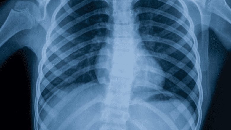
X-rays are helpful in diagnosing spinal arthritis.
The doctor may order an X-ray to see if there is joint damage and how much joint damage has occurred. The X-ray can show cartilage loss, compression fractures, and the presence and location of bone spurs. X-rays are also useful in helping to exclude other causes of pain and to better inform possible considerations for referral to a specialist.
However, it is important to keep in mind that what shows up in an X-ray may not correlate to the presence or absence of osteoarthritis and associated pain. For example;
- Most people over age 60 have degenerative changes in their spine consistent with osteoarthritis, but for perhaps 85% of them there is no pain or stiffness.
- Conversely, an X-ray conducted during the early stages of osteoarthritis may not yet show any visible damage to the joints, but the patient may have symptoms.
For all these reasons, the clinical history and physical examination are essential to arriving at an accurate clinical diagnosis and plan of treatment.
CT Scan
A CT scan may be used to better show the adequacy of the spinal canal and surrounding structures.
A CT scan may also include myelography, which involves an x-ray contrast dye that is injected into the spinal column to show issues such as a bulging disc or bone spur possibly pressing on the spinal cord or nerves.
MRI Scan
The MRI, or magnetic resonance imaging scan, is a sophisticated imaging method that can show detailed images of the spinal cord, nerve roots, discs, ligaments, and surrounding tissues and spaces.
Most MRI scans require the patient to lie flat in a tube for about 40 minutes. Open frame, and even standup, MRI scanners do exist and may be appropriate for patients having claustrophobia (fear of tight spaces).
MRI scans can be adjusted to show details of tissues, such as water content in the tissue, which may be important in determining disc degeneration, infections, or tumors.
Serious Conditions Related to Lower Back Pain
Severe numbness or weakness in one or both legs requires immediate medical attention
For symptoms of lower back pain PLUS any of the following red flags then a medical evaluation is an emergency, and should be done same day:
- History of cancer, or unexplained weight loss
- Symptoms of infection, such as fever, shakes, chills
- Numbness in the perineum (genital areas) and urinary problems
- Recent fall or trauma that may have caused spine fracture
- Severe numbness or weakness in one or both legs
See When Back Pain May Be a Medical Emergency
The evaluation usually consists of a discussion of symptoms and a detailed medical history, a physical examination, and X-rays of the low back.
Other tests (blood tests, MRI or CT scans) may be performed if there are red flags, or to confirm or exclude the presence other rare conditions that can cause similar symptoms, such as a spinal tumor, infection, fracture, or other types of arthritis. MRI and CT scans are not routinely needed in the initial evaluation of low back pain. Studies have shown patients who get MRI imaging in the early stages actually tend to do worse due to over-treatment — since so much of the general population has irregularities in their spine, early MRIs may lead doctors to incorrectly associate an irregular finding as the source of the patient’s pain.
The goal of all diagnostic studies is to discover patterns or confirmations between the various tests that point to a clear diagnosis among various possible ones.
See Introduction to Diagnostic Studies for Back and Neck Pain
The key is to diagnose the condition causing the patient's pain and disability, which includes putting all the pieces of the puzzle together and does not rely on diagnostic tests alone.


